
Figure 2.
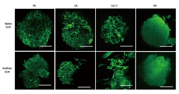
Three-dimensional extracellular matrix scaffolds derived from pluripotent stem cell aggregates. Confocal images of fibronectin (FN), laminin (LN), Collagen IV (Col IV), and vitronectin (VN) expression pre- and post-decellularization [acellular extracellular matrix (ECM) and native ECM, respectively]. Scale bar: 100 μm. For native Col IV, scale bar: 50 μm. The ECM scaffolds can be used for neural differentiation. Images are adapted from Sart et al[60].