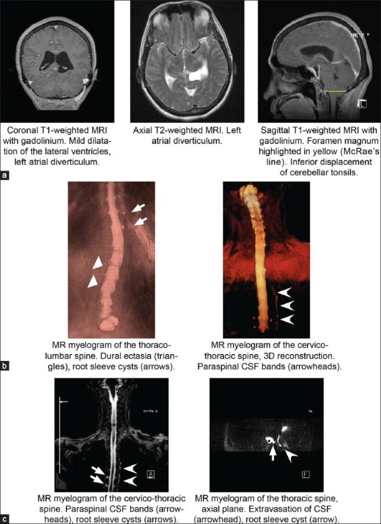
Figure 1.

Diagnostic imaging. (a) Coronal and sagittal T1 with gadolinium and axial T2 MRI demonstrating mild dilatation of the lateral ventricles, left atrial diverticulum, and inferior displacement of the cerebellar tonsils (McRae's line in yellow). (b) MR myelography of the thoraco-lumbar and cervico-thoracic spine with 3D reconstruction demonstrating lumbar DE (triangles), root sleeve cysts (arrows), and paraspinal CSF bands (arrowheads). (c) MR myelography of the cervico-thoracic spine demonstrating root sleeve cysts (arrows) and extravasation of CSF with paraspinal CSF band (arrowheads)
