Figure 1.

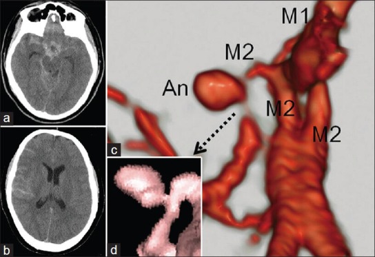
Computed tomography (CT) of the patient at admission showed a thick subarachnoid hemorrhage (SAH) predominantly in the basal cistern and right Sylvian fissure (a). The SAH was spreading to the peripheral subarachnoid space and the brain seemed really tight (b). Three-dimensional CT angiography revealed an aneurysm arising from a distal point of the right middle cerebral artery bifurcation (c). Magnetic resonance (MR) angiography shows a clear image of the stalk-like and narrow aneurysm neck (d)
