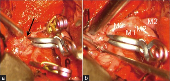
Figure 2.

Operative pictures of the aneurysm clipping. (a) The aneurysm was buried into the right frontal lobe (arrow). With parent artery trapping, the aneurysm was tentatively clipped. (b) Final view of the clipping. The aneurysm was carefully resected. There was no obvious arterial wall (asterisk). The aneurysm was located distal to the right middle cerebral artery bifurcation without an adjacent artery
