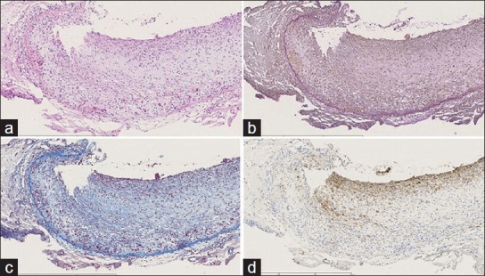
Figure 3.

Histopathological studies of the surgical specimen. (a) Hematoxylin and eosin (H and E) staining. The upper side is the vascular lumen. Atherosclerotic change was not observed. (b) Elastica van Gieson (EVG) staining shows thickened intima and mild invasion of inflammatory cells. (c) Masson's trichrome staining. (d) SMA (smooth muscle actin) immunohistochemistry. Staining reveals lack of smooth muscle cells in the tunica media. Scale bar: 600 μm
