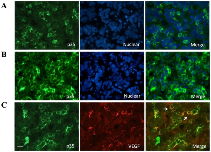
Figure 2.
A, Presence of p35 in normal human pituitary. Immunofluorescence double-staining for p35 (green) and nuclear (blue) was performed; B, Presence of P35 in pituitary adenomas. Immunofluorescence double-staining for P35 (green) and nuclear (blue) was performed; C, Colocalization of p35 and and VEGF in the normal human pituitary gland. Immunofluorescence double-staining for p35 (green) and VEGF (red) was performed. Colocalization of both is indicated by filled arrowheads. Data are representative of at least three different human adult pituitary samples and at least three independent experiments. Scale bar, 20μm.

