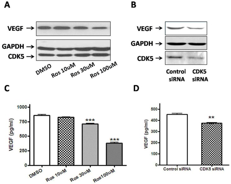
Figure 5.
A, Pretreatment with roscovitine significantly decreased VEGF expression in GH3 cells. Blots were reprobed with anti- GAPDH antibody to en sure equal loading; 5B, Effect of Cdk5 siRNA on VEGF expression in GH3 cells. Blots were reprobed with anti- GAPDH antibody to en sure equal loading; 5C, Quantification of the level of VEGF secretion from vehicle or roscovitine-treated GH3 cells. Reduction of VEGF secretion were detected in roscovitine-treated GH3 cells compared with vehicle group; 5D, Quantification of the level of VEGF secretion from control siRNA or CDK5 siRNA-transfected GH3 cells. Reduction of VEGF secretion were detected in CDK5 siRNA-transfected GH3 cells compared with vehicle group. VEGF secretion was quantified as by ELISA as described above. Data represents means ± SEM of three independent experiments. one-way ANOVA was performed to determine the significance (**p < 0.01 and ***p < 0.001).

