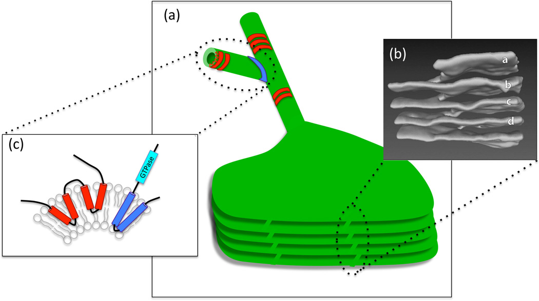
Figure 1.
Diverse membrane structures in the ER. (a) The ER is an interconnected network of composed of branched tubules and sheets, some of which can form stacks, as shown in the illustration. ER tubules are stabilized by the oligomerization of proteins such as reticulons and DP1/Yop1/REEps (in red), while 3-way junctions are mediated by proteins such as atlastins (in blue). (b) The structure of reticulons and atlastin. Membrane curvature is induced by the insertion of protein "wedges" (two in the case of reticulons and one in the case of atlastin) that traverse only one lipid bilayer, forcing the membrane to curve. Atlastin has an in addition GTPase activity that is necessary for fusing membranes and generating 3-way junctions. (c) Helicoidal membrane structure in stacked ER sheets. A 3D reconstruction of a region of stacked ER sheets from an acinar cell of a mouse salivary gland. The letters (a through d) mark the ER sheets that are connected through a helicoidal structure (to the left). From Terasaki et al. Cell 154, 285–296, 2013.

