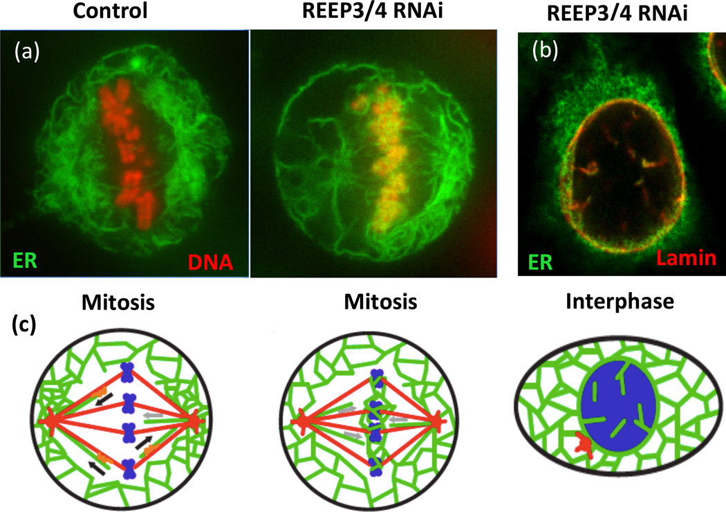
Figure 2.
Depletion of REEP3/4 causes accumulation of ER on mitotic chromosomes and leads to intranuclear membranes and lamina. A. HeLa cells expressing RFP-histone (red) to label the DNA and GFP-Sec61 (green) to mark the ER were subjected to control or REEP3/4 RNAi and imaged during mitosis. Note the colocalization of ER and mitotic chromosomes in the absence of REEP3/4. (b) An interphase REEP3/4 RNAi-treated HeLa cell expressing GFP-Sec61 (green) was fixed and stained for nuclear lamin B1 (red). Both NE markers are aberrantly localized to structures inside the nucleus. (c) Schematic of phenotypes with microtubules in red, DNA in blue and ER in green. Adapted from Schlaitz et al. Dev. Cell 26, 316–323, 2013. Images courtesy of Anne-Lore Schlaitz and Rebecca Heald.

