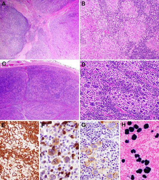
Figure 3.
Unusual morphological findings in some examples of EBV-positive NLPHL . Case 3, (also illustrated in Figure 2) demonstrates capsular fibrosis with focal fibrous septa (A). Case 6 (also illustrated in Figure 1) exhibits a prominent granulomatous reaction with focal areas of necrosis (B). In case 10 the low power nodular appearance is typical (C). However, the LP cells are larger than expected with more pleomorphic and polylobated nuclei arranged in clusters (D). They are strongly positive for CD20 (E), which also highlights the small B cells in the background. They are weakly positive for CD79a (F) and CD30 (G). EBER was positive (H).

