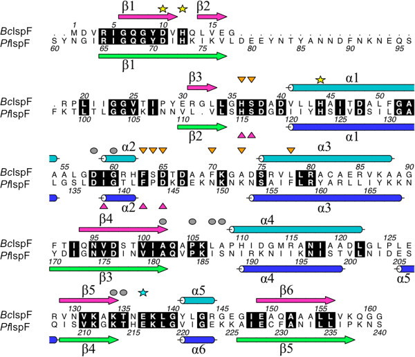
Figure 3.
Structure-based sequence alignment of BcIspF and PfIspF. The alignment has been annotated with elements of secondary structure. Strictly conserved residues are highlighted in black. Three residues that coordinate Zn2+ are marked with a yellow star. Residues that bind CDP or CMP 1, are marked with a grey disk and those that bind CMP 2 with an orange triangle. Residues that bind MEcPP in EcIspF [20] are marked with a magenta triangle. A conserved glutamate that binds either Mg2+ or Mn2+ in EcIspF structures is highlighted with a cyan star. This figure was prepared using ALINE[29].

