Figure 7.

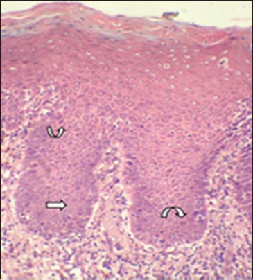
Photomicrograph showing basilar hyperplasia (straight arrow) and hyperchromtic nuclei in group3 (curved arrow). (H&E stain, ×200)

Photomicrograph showing basilar hyperplasia (straight arrow) and hyperchromtic nuclei in group3 (curved arrow). (H&E stain, ×200)