Figure 5.

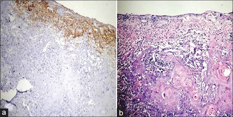
(a) Histopathological image shows well differentiated oral squamous cell carcinoma: VEGF staining intensity 2× (IHC stain, ×100) (b) Histopathological image shows well differentiated oral squamous cell carcinoma.(H&E stain, ×100)

(a) Histopathological image shows well differentiated oral squamous cell carcinoma: VEGF staining intensity 2× (IHC stain, ×100) (b) Histopathological image shows well differentiated oral squamous cell carcinoma.(H&E stain, ×100)