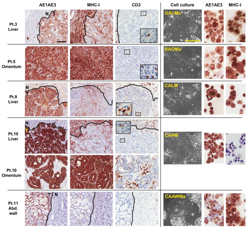
Fig. 1. Cancer cell lines established from gastrointestinal cancer metastases with heterogeneous MHC-I expression and weak T-cell infiltration.
Immunohistochemical staining of paraffin-embedded gastrointestinal cancer metastases (left) in five patients (Pt.) and representative corresponding cancer cell lines (right). Pt. 3, gastric adenocarcinoma liver metastasis, adjacent normal liver, and derived cancer cell line (GALMa). Pt. 5, omental metastasis from an intrahepatic bile duct adenocarcinoma and derived cancer cell line (BAOMa). Pt. 9, colon adenocarcinoma liver metastasis, adjacent liver, and derived cancer cell line (CALM). Patient 10, colon adenocarcinoma liver metastasis, adjacent liver, and omental metastasis, with derived cancer cell line (CAPM). Patient 11, abdominal wall metastasis from a mucinous colon adenocarcinoma, adjacent stroma, and derived cancer cell line (CAAWMa). All epithelial derived-cancer cells are stained by the pan-cytokeratin marker AE1AE3 which delineate tumor (T) from the adjacent normal tissues (N). MHC class I stains all nucleated cells, with weak and heterogeneous expression found in liver metastases in vivo, however most derived cancer cell line express MHC class I in culture except CAPM. Tumor-infiltrating lymphocytes are revealed by CD3 and T-cell marker, and in all cases represent less than 5% of cells in tumor mass.
Representative fields selected from whole slide scan, measure bar = 100 um.

