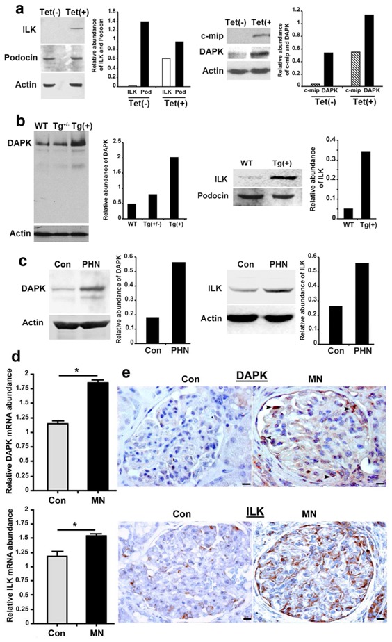
Figure 5. c-mip induces in vitro and in vivo an upregulation of ILK and DAPK.

(a) Representative Western blots of ILK, podocin, c-mip and DAPK on protein lysates from non-induced [Tet(-)] and induced [Tet(+)] stable transfectant podocytes. (b) Representative Western blots of DAPK and ILK on glomerular lysates from wild-type and transgenic mice. (c) Representative Western blots of DAPK and ILK on glomerular lysates from PHN and control rat kidneys (Con). (d) Quantitative RT-PCR of laser microdissected glomeruli from MN kidney biopsy specimens (n=5) and control kidneys (n=5); * P <0.05, Mann-Whitney test. (e) Representative immunohistochemical analysis of DAPK and ILK in serial sections of kidney biopsy specimens from patients with MN and control human kidney (Con). Scale bars, 20 μM
