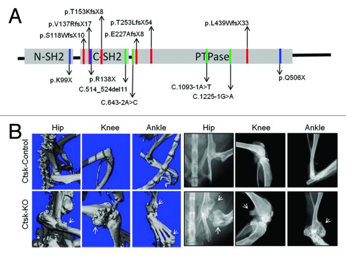
Figure 1. (A) PTPN11 mutations identified in MC patients. The locations of MC mutations in the corresponding SHP2 structure are indicated by red, blue and green colored lines, representing frameshift, nonsense and splice-site mutations, respectively. Predicted protein changes are indicated with arrows. Please see references 1 and 2 for original references. (B) Micro-CT and Faxitron images demonstrate the existence of osteochondromas and enchondromas (arrows) at hip, knee and ankles of 12-week-old mice lacking SHP2 in cathepsin K-expressing cells (CtskCre;Ptpn11fl/fl, Ctsk-KO) but not its littermate controls (CtskCre;Ptpn11fl/+, Ctsk-Control).
