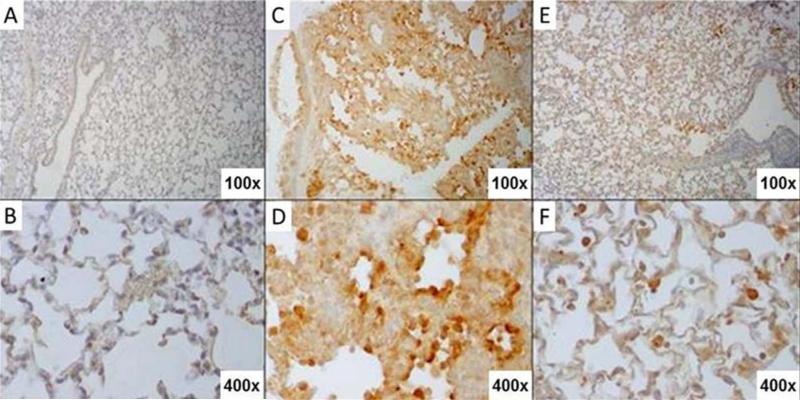
Fig. 5. EUK-207 treatment reduces oxidative damage during 1918 influenza virus infection.
Immunohistochemistry for oxidation marker 8-Oxo-2'-deoxyguanosine (8-OHdG). (A) Staining of mock infected, untreated mouse lung. (B) Close up of the alveolar epithelium from mock infected, untreated mouse lung from panel A. (C) Staining of vehicle-treated 1918 influenza virus-infected mouse lung showing prominent 8-OHdG staining in bronchial epithelium as well as alveolar epithelium. (D) Close up view of alveolar epithelium from panel C showing 8-OHdG staining. Prominently staining alveolar epithelial cells showing type II hyperplasia are shown also with staining in inflammatory cells in interstitial infiltrates. (E) Staining of EUK-207-treated 1918 influenza virus-infected mouse lung showing marked reduction in 8-OHdG staining in bronchial epithelium as well as alveolar epithelium as compared to the lung from the vehicle-treated animal in panel C. (F) Close up view of alveolar epithelium from panel E showing staining in alveolar macrophages and a marked reduction in 8OH-dG staining in alveolar epithelium. Original magnifications for each image are noted in their bottom right hand corners.

