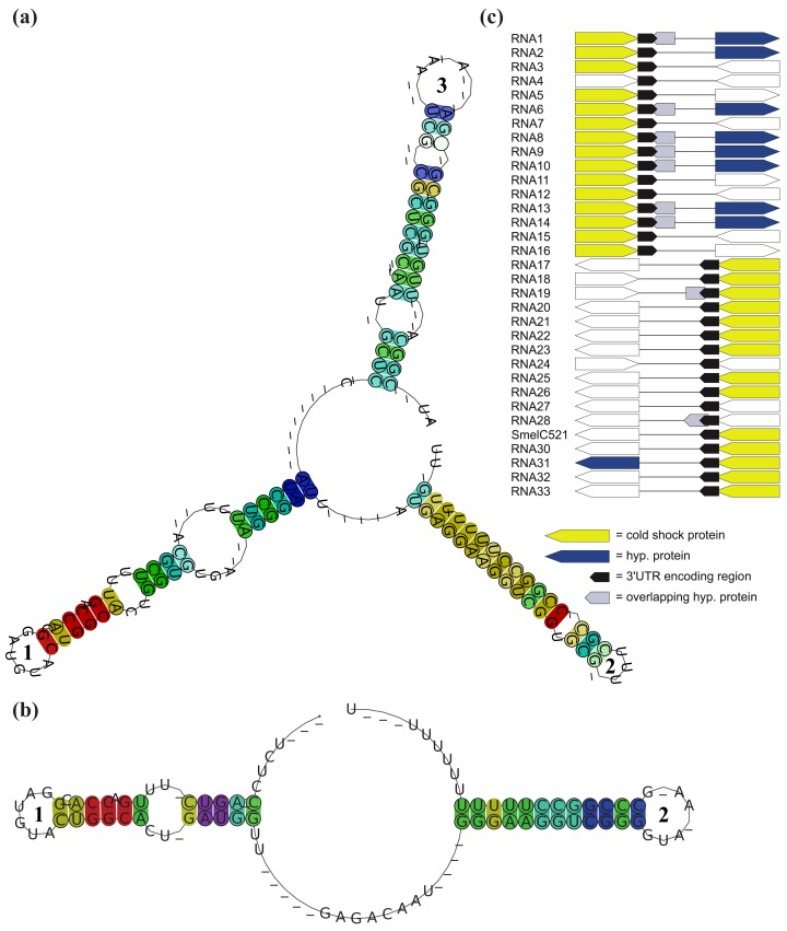
Figure 4.
Structural comparison between RFMSmelB053 and related 3′-UTRs. (a) Consensus secondary structure of RFMSmelB053 and (b) related 3′-UTRs; Base pairs in (a) and (b) are colored according to the Vienna RNA conservation coloring scheme [65]. Colors indicate the number of nucleotide combinations, out of the six possible base pairs, in the underlying alignment that are involved in forming predicted base-pairs (red = 1, yellow = 2, green = 3, cyan = 4, blue = 5, purple = 6). Pale colors are used for the case that some sRNAs do not form a base-pair; (c) Associated proteins of 3′-UTRs. Arrows indicate the orientation of each gene, identical colors indicate homologous genes. Non-colored arrows denote non-homologous genes (genes are not shown to scale).

