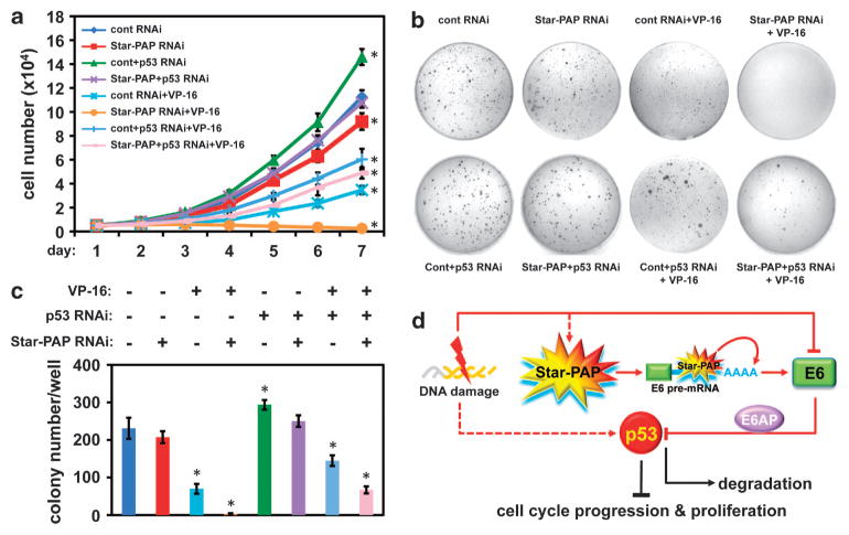
Figure 3.
Star-PAP-mediated p53 expression regulates cervical cancer cell proliferation. (a) Analysis of HeLa cell proliferation34 after Star-PAP or/and p53 knockdown in the presence or absence of contiguous low dose of VP-16 (2 μM) treatment for a period of time as indicated. Error bars represent s.e.m. of three individual experiments with triplicate for each experimental condition. For statistics, individual experimental conditions are compared with control group. *P≤0.05 (t-test). p53 small interfering RNA (siRNA) 5′-CCGUCCCAAGCAAUGGAUGAUUUGA-3′. (b) Soft agar colony formation assay using HeLa cells treated under the same conditions as described above. A representative batch of experiment is displayed, and the colony numbers of each experimental condition were quantified and the means of the colony numbers from three independent experimental runs were used for plotting (c). Error bars represent s.e.m. of three individual experiments. For statistics, individual experimental conditions are compared with control group. *P≤0.05 (t-test). This assay was carried out in 35 mm cell culture dishes, where three layers of soft agar (Fisher Scientific, #BP1423, Pittsburgh, PA, USA; bottom 0.6%, middle 0.4% and top 0.6%) were plated individually (0.5 ml liquefied agar at 40 °C for each layer) with the middle layer containing 104 cells per dish. After plating the sandwich agar and the agar solidified, 1 × Dulbecco’s modified Eagle’s medium containing 10% fetal bovine serum and penicillin/streptomycin (50 U/ml) was overlaid on top and the cultures were grown at 37 °C in 5% CO2 for 4 weeks. The colonies were stained with Giemsa Stain (Sigma-Aldrich, #GS500, St Louis, MO, USA) and visible colonies were counted and quantified. (d) Model of the Star-PAP-regulated p53 expression in the control of cell proliferation in response to DNA damage signal.

