Figure 1.
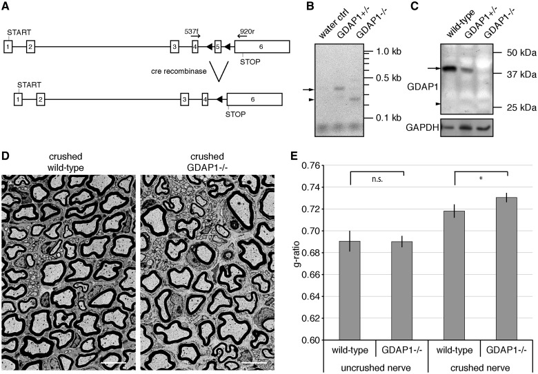
Gdap1−/− animals display hypomyelination after nerve crush. (A) The ablation of exon 5 in the Gdap1 locus generates a premature stop codon in exon 6 as a result of a frame shift (triangles: introduced loxP sites; START: translation start codon; STOP: translational stop codon; arrows indicate primer pairs used for reverse transcriptase-PCR). (B) Reverse transcriptase-PCR demonstrates the trans-splicing from exon 4 to 6 in Gdap1−/− animals. The sizes of the predicted PCR products for the wild-type transcript (arrow; 383 bp) and the trans-spliced transcript (arrow head; 269 bp) are indicated. (C) Loss of GDAP1 protein in mutant mice (arrow: wild-type protein; arrowhead: size of the predicted GDAP1 K193fsX219 protein: note that the antiserum detects the amino-terminus of GDAP1). Glyceraldehyde 3-phosphate dehydrogenase (GAPDH) served as loading control. (D) Representative ultrathin sections of unilaterally crush-lesioned sciatic nerves of 4-month-old wild-type and Gdap1−/− mice analysed by transmission electron microscopy. Two month post crush sections were obtained 3 mm distal to the site of injury. Scale bar = 5 µm. (E) Quantitative morphological analysis of sciatic nerve sections. One hundred randomly selected axons per crushed and uncrushed nerve were measured per animal. Values represent mean and standard error of three animals per genotype: n.s. = not significant, *P < 0.05.

