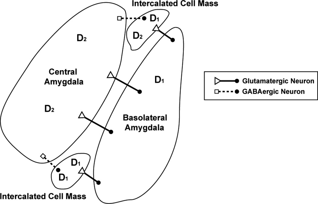
Figure 3. Distribution of dopamine receptors in the amygdala.
Neurons in the central amygdala primarily express dopamine D2 receptors (Weiner et al., 1991). Blockade of these receptors may lead to impaired fear conditioning (Guaracci et al., 2000) or prevent the animal from generating appropriate fear or reward-related behaviors. The central amygdala receives glutamatergic input (open triangles) from the basolateral amygdala (cell body represented by filled circles). The basolateral amygdala (BLA) coordinates activity from the nucleus accumbens, prefrontal cortex, and many other regions to generate and retrieve CS-US associations. Dopamine D1 receptors are primarily expressed in the BLA and intercalated cell masses of the amygdala (ITC). Antagonism of D1 receptors in the basolateral amygdala impairs acquisition of fear extinction (Hikind & Maroun, 2008). Glutamatergic neurons from the BLA project to ITC and GABAergic ITC neurons inhibit central amygdala activity (output represented by open squares). The activation of GABAergic ITC neurons mediated by the basolateral amygdala and the infralimbic region of the prefrontal cortex is critical to extinction of fear behaviors and inhibitory control of the central amygdala (Busti et al., 2011). In addition to high D1 receptor expression, ITC express D2 receptors at low levels (Weiner et al., 1991). In summary, the release of dopamine in the amygdala can have complex effects on behavior due to the distribution of dopamine receptors and the dopamine receptor subfamilies expressed in this region.

