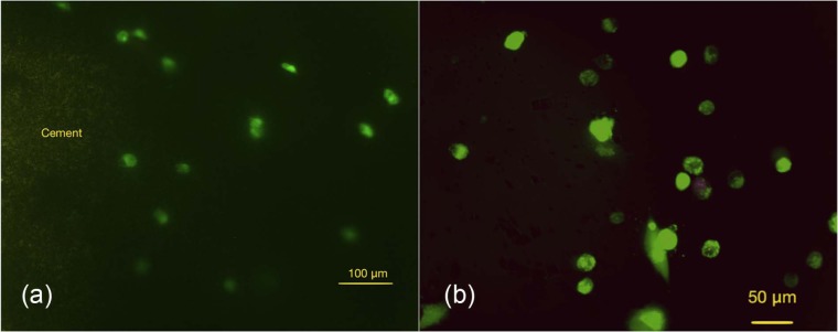
Figure 10.

Photomicrographs for live and dead staining (calcein-AM and ethidium homodimer) for rMSCs in collagen after 24 h of cell culture: (a) presence of live rMSCs around the cement (Cerament Spine Support; green) after 24 h of cell culture (scale bare = 100 µm); (b) live cells inside a rat-tail collagen without cement (control) after 24 h of cell culture (scale bar = 50 µm). Result confirms presence of live cell evenly distributed around the injected cement.
calcein-AM: acetomethoxy derivate of calcein; rMSCs: rabbit mesenchymal stromal cells.
