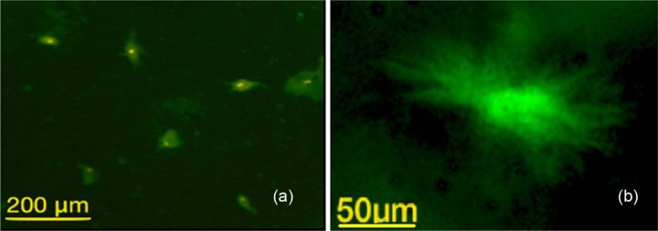
Figure 11.

Photomicrography showing immunofluorescent stain for rMSCs showing positive staining to OCN, an antibody: (a) presence of multiple rMSCs with positive intake to OCN antibodies (scale bar = 200 µm); (b) on-cell ith higher magnification (scale bar = 50 µm), which indicates the pre-osteoblast differentiation of the MSCs after seeding into the collagen around the CS/HA cement.
OCN: osteocalcin; CS/HA: calcium sulphate/hydroxyapatite; rMSCs: rabbit mesenchymal stromal cells.
