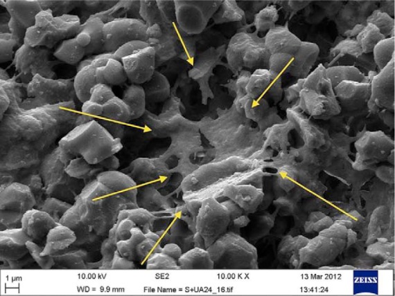
Figure 6.

Scanning electron microscopy image showing rMSC (arrows) at day 1 of cell culture. The cell is covering micropore and is adherent onto the surface of the Cerament Spine Support cement (scale bar = 1 µm).
rMSCs: rabbit mesenchymal stromal cells.
