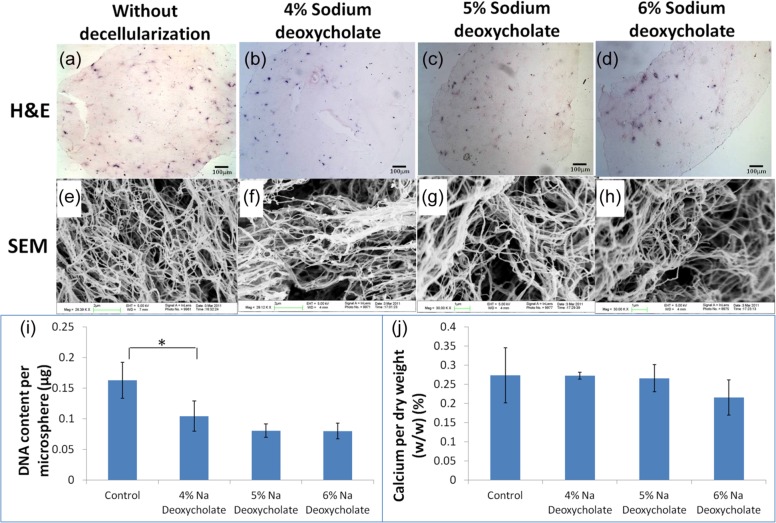
Figure 2.
Decellularization of osteogenic differentiated MSC–collagen constructs with detergent at different dosages. (a–d) Routine H&E staining (scale bar: 100 µm); (e–h) SEM images; (a and e) without decellularization; (b and f) decellularized with 4% sodium deoxycholate; (c and g) decellularized with 5% sodium deoxycholate; (d and h) decellularized with 6% deoxycholate; (i) DNA content after decellularization (*statistical significant difference: p = 0.05); (j) calcium content per dry weight after decellularization (n = 3, each with duplicates).
MSC: mesenchymal stem/stromal cell; H&E: hematoxylin and eosin; SEM: scanning electron microscope.

