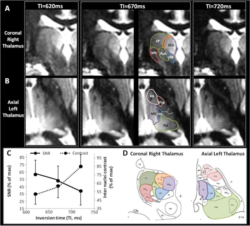
Figure 5. Fine optimization of TI.
Different TI were tested around the WM nulling point for TS=6000ms. Representative coronal images of the right thalamus (A) and axial images of the left thalamus (B) at three different TIs close to the WM nulling point, as noted. Other parameters fixed at TS=6000ms, N=200, BW=15KHz, TR=10.2ms, TE=4.4ms, α =4°, resolution=1mm3, ARC acceleration factor=2.5, NEX=1, scan time=8.2min. TI=670ms is shown without and with annotations. The average SNR and the average contrast between multiple pairs of adjacent nuclei were measured for each condition and plotted in (C). The corresponding histological plates from the Morel atlas (Morel et al., 1997) are shown for anatomic correspondence in (D).
From the simulations (see Figure 2A and 2B), the thalamic signal was expected to decrease with longer TI around the WM null regime, but the intra-thalamic contrast (relative difference between the signal curves for nuclei with low and higher T1 values) was expected to increase for longer TI around the WM null regime. Experimentally, the trade-off between thalamic signal and contrast along TI was confirmed (C). Several nuclei were better delineated at TI 670ms. For example, on the coronal view (A and D) the hyperintensity of LP (green) and MD (purple) was distinct from the more hypointense VPL (red), CM (blue) and CL (orange). Also, a thin hypointense border allowed a clear delineation of the PuA (green) that was not possible at TI=620ms due to insufficient contrast, nor at TI=720ms, due to insufficient signal. Similarly, on axial views (B and D), the thin hypointense border delineating VA (pink) was better seen at TI=670ms. Same observation for the hypointense triangle corresponding to VLa (dark pink) seen at TI=670ms (and to a lesser extent at TI=620ms) but not individualized at TI=720ms due to the loss of signal from the adjacent posterior nuclei. Some subtle boundaries were also made out (dashed lines) and will be better emphasized by scanning without parallel imaging.
The WM null regime at TS=6000ms and TI=670ms was preferred for the next steps of the optimization for thalamus segmentation.

