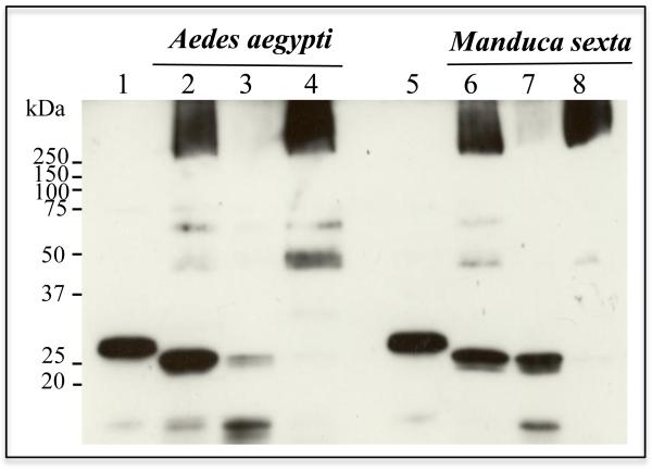
Figure 5.
Cyt1Aa oligomers insert into BBMV of Aedes aegypti and Manduca sexta. Cyt1Aa protoxin was activated with proteinase K in the presence of BBMV from both insects, finally the BBMV membranes were separated by centrifugation. The membrane pellets and the supernatants were loaded on SDS-PAGE and Cyt1Aa was revealed by western blot by anti-Cyt1Aa antibody. Lanes 2 to 4 correspond to A. aegypti BBMV while lanes 6 to 8 to M. sexta BBMV. Lanes 1 and 5 are Cyt1Aa protoxin samples, lanes 2 and 6 the correspond to the BBMV samples without separation by centrifugation, lanes 3 and 7 are the supernatants after separation of the membranes by centrifugation and lanes 4 and 8 are the BBMV membrane pellets after separation by centrifugation.

