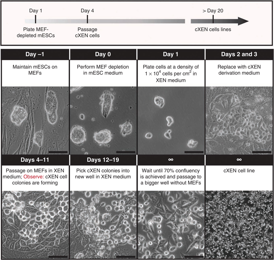
Figure 5.
Timeline for cXEN cell derivation from mouse ES cells. Protocol for the conversion of ES cells to cXEN cells using growth factors. Day − 1: ES cells are maintained in ES medium on MEFs. Day 0: ES cells are passaged onto a pregelatinized plate for MEF depletion. Day 1: ES cells are enzymatically passaged with 0.05% (wt/vol) trypsin and plated at a density of 1 × 10⁴ cells per cm2 in standard XEN medium. Days 2 and 3: the medium is replaced with cXEN derivation medium daily (0.01–10 µM retinoic acid plus 10 ng ml− 1 activin A). Days 4–11: cells are enzymatically passaged onto MEFs in XEN medium. The medium is replaced every day or every other day depending on confluency. XEN-like cells with stellate and refractile morphology will emerge within ~5 d. Days 12–19: XEN-like cells are picked manually and placed in a MEF-coated or pregelatinized plate in XEN medium. They are passaged two more times onto pregelatinized plates and the cXEN cell line is frozen. Scale bars, 100 µm.

