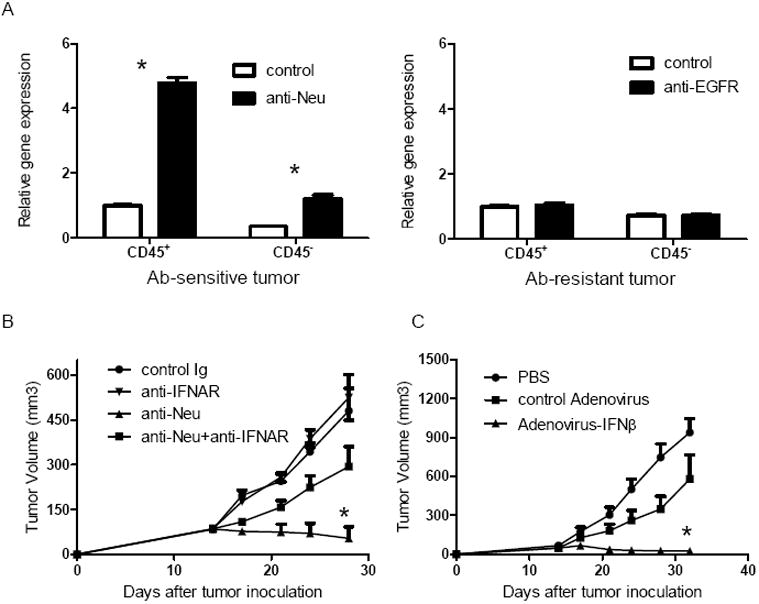
Figure 1. Type I interferons are induced and necessary during antibody-mediated tumor regression.

A) WT BALB/c mice (n=4/group) were injected subcutaneously with 5×10⁵ TUBO cells, then 100 μg of anti-Neu (left panel) or control IgG was administered on day 14. WT B6 mice (n=4/group) were injected subcutaneously with 7×10⁵ B16-EGFR cells, then 100 μg of anti-EGFR (right panel) or control IgG was administered on day 14. Four days after the treatment, tumor was digested and sorted to CD45+ and CD45- population. Quantitive real-time PCR was performed to detect the expression of mRNA levels of mIFNα5. Mean + SD are shown. B) WT BALB/c mice (n=5/group) were injected subcutaneously with 5×10⁵ TUBO-EGFR cells, then 100 μg of anti-Neu was administered on days 14 and 21. 200ug of anti-IFNAR or control Ig was intratumorally administrated on the same days. The growth of tumor was measured and compared twice a week. Mean + SEM are shown. C) WT B6 mice (n=5/group) were injected subcutaneously with 5×10⁵ B16-EGFR cells, then 2×10⁹ Adenovirus-IFNβ or control virus was intratumorally administrated on days 14 and 21. The growth of tumor was measured and compared twice a week. *, Mean + SEM are shown. p < 0.05 compared with control group. One representative experiment of three is depicted. See also Figure S1.
