
Figure 9.
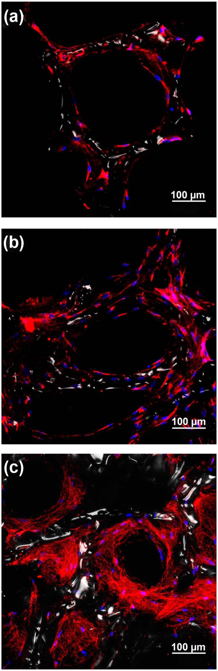
Confocal laser scanning microscopy visualization of type I collagen deposition in alginate-coated TiO2 scaffolds with or without simvastatin (SIM). Fluorescence immunocytochemical analysis of type I collagen in primary human osteoblasts cultured on alginate-coated TiO2 scaffolds. Type I collagen is detected in the majority of the cells cultured on scaffolds with (a) 10 nM SIM, (b) 10 µM SIM and (c) without SIM. Extracellular collagen fibrils are only seen in scaffold (c) without SIM. Type I collagen (red), DNA (blue), TiO2 scaffold surface (white).