Figure 2.
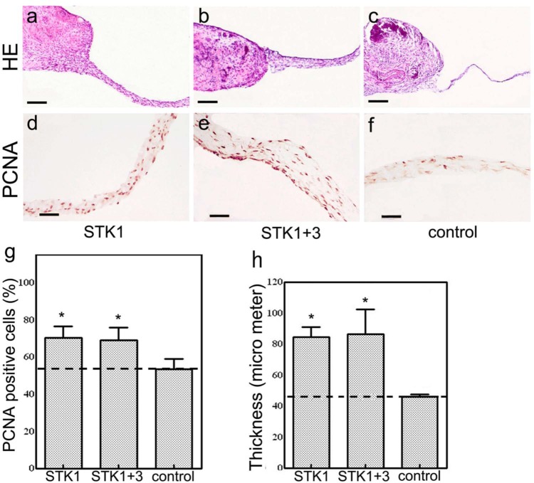
(a–c) Histology of CPSs (hematoxylin and eosin staining). CPSs expanded with 1% HS-STK1 and 1% HS-STK1+3 exhibiting thicker outgrowth of the peripheral region formed by extracellular matrix–rich multilayered cells than 10% FBS-M199. (d–f) Immunohistochemical observations of PCNA-positive cells. (g) PCNA-positive cells were notably increased at the peripheral region of the CPSs expanded with 1% HS-STK1 and 1% HS-STK1+3. (h) Measurement of CPS thickness. (a–c) bar, 200 µm and (d–f) bar, 50 µm.
CPS: cultured periosteal sheet; HS: human serum supplemented; FBS: fetal bovine serum; PCNA: proliferating cell nuclear antigen.
n = 3, *p < 0.05, **p < 0.01, compared with controls.

