Figure 6.
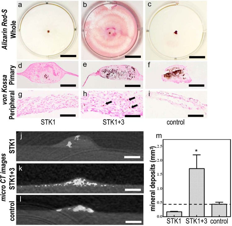
(a–i) Alizarin red-S and von Kossa staining of CPSs. (a–f) Under all conditions, the dye-affinity of Alizarin red-S and von Kossa was confirmed in the primary periosteal fragment, (b and h: arrows), while CPSs expanded with 1% HS-STK1+3 exhibited only enlargement of satiability extending to the peripheral region. (j–l) Micro CT images in the CPSs. (j and l) In CPSs of 1% HS-STK1 and 10% FBS-M199, mineral deposition was limited to primary periosteal fragments. (k and m) A CPS of 1% HS-STK1+3 exhibited mineral deposition occurring over a wide range of CPSs and a remarkable increase in total mineralized volume. (a–c). Primary: region of the primary periosteal fragment and peripheral: area of the peripheral outgrowth of the periosteal cells Bar, 10 mm; (d–f) bar 400 µm; and (g–i) bar, 100 µm.
CPS: cultured periosteal sheet; HS: human serum supplemented; CT: computed tomography; FBS: fetal bovine serum.
n = 3, *p < 0.05, compared with the controls.

