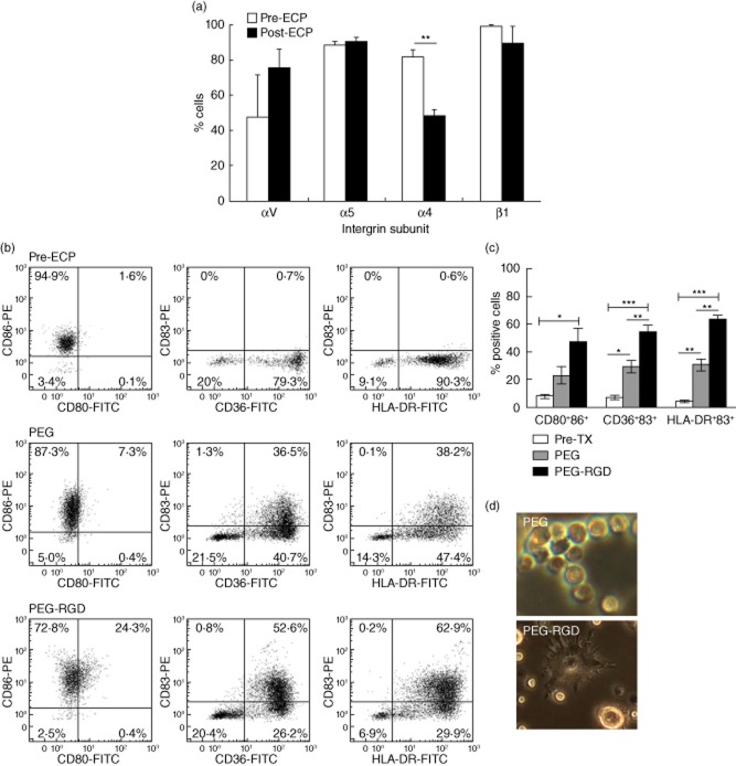
Figure 3.
Monocyte-to-dendritic cell (DC) conversion is stimulated by arginine–glycine–aspartic (RGD)-conjugated polyethylene glycol (PEG) hydrogels. (a) Modulation of integrin subunit expression after overnight incubation of extracorporeal photochemotherapy (ECP)-treated samples (post-ECP, n = 3) in comparison to peripheral blood monocytes (pre-ECP, n = 4). **P < 0·01 as confirmed by Student's t-test. (b) Purified monocytes were cultured overnight on PEG hydrogels conjugated with RGD (PEG–RGD) or unmodified PEG hydrogels (PEG). Dendritic cells were identified by co-expression of membrane CD80 and CD86 and membrane staining for CD36 or human leucocyte antigen D-related (HLA-DR) combined with cytoplasmic reactivity with CD83. Representative two-colour histograms from freshly isolated monocytes cultivated on PEG or PEG–RGD hydrogels. (c) Average fraction of DC obtained using PEG–RGD or PEG hydrogels. *P < 0·05; **P < 0·01; ***P < 0·0001 as confirmed by analysis of variance (anova) and paired Student's t-test. (d) Phase-contrast microscopy demonstrates poor dendrite formation on ECP differentiated DC on PEG with improved morphology on PEG–RGD.

