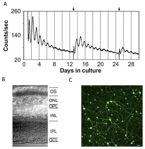
Figure 3. Bioluminescence Rhythms from mPer2Luc Mouse Retinal Explants.
(A) Long-term culture of an intact mouse retinal explant showing persistent circadian rhythms in PER2::LUC expression. Arrows indicate times of media changes. (B) Representative DIC image of vertical retinal sections. Retinal explants were cultured, and vertical retinal slices were cut with a tissue slicer at 150 μm. GCL, ganglion cell layer; INL, inner nuclear layer; IPL, inner plexiform layer; ONL, outer nuclear layer; OPL, outer plexiform layer; OS, photoreceptor outer segments. (C) Flat-mount view showing tyrosine hydroxylase immunoreactivity in cultured retinal explants. The immunoreactive cells with relatively large somata and two to three thick primary processes that arise from the cell body are dopaminergic amacrine cells, whereas the immunoreactive cells with relatively small cell body and very few processes are type 2 catecholamine amacrine cells. (adapted from Ruan et al., , Plos Biol. 2008).

