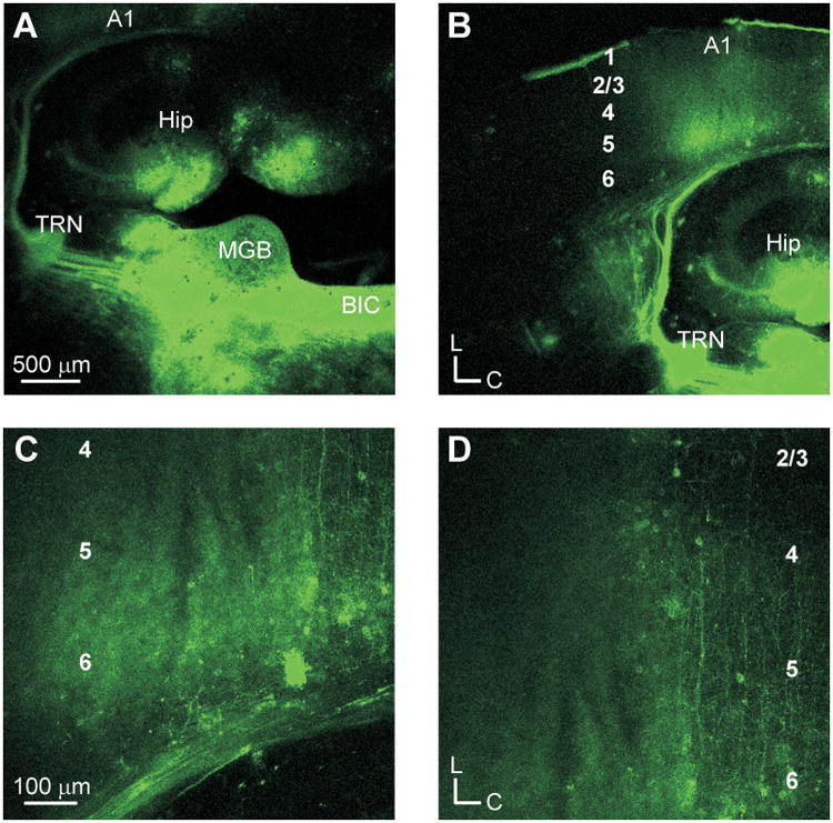
Fig. 1.

Thalamocortical projections in the auditory slice preparation. (A) Placement of DiI crystals in the medial geniculate body (MGB). Labeled fibers traverse toward the thalamic reticular nucleus (TRN) continuing onward towards the primary auditory cortex (A1). (B) Thalamocortical fiber terminations in A1. (C) Labeled axonal fibers and retrogradely labeled cells in layer 6. (D) Labeled fibers extending to layer 4.
