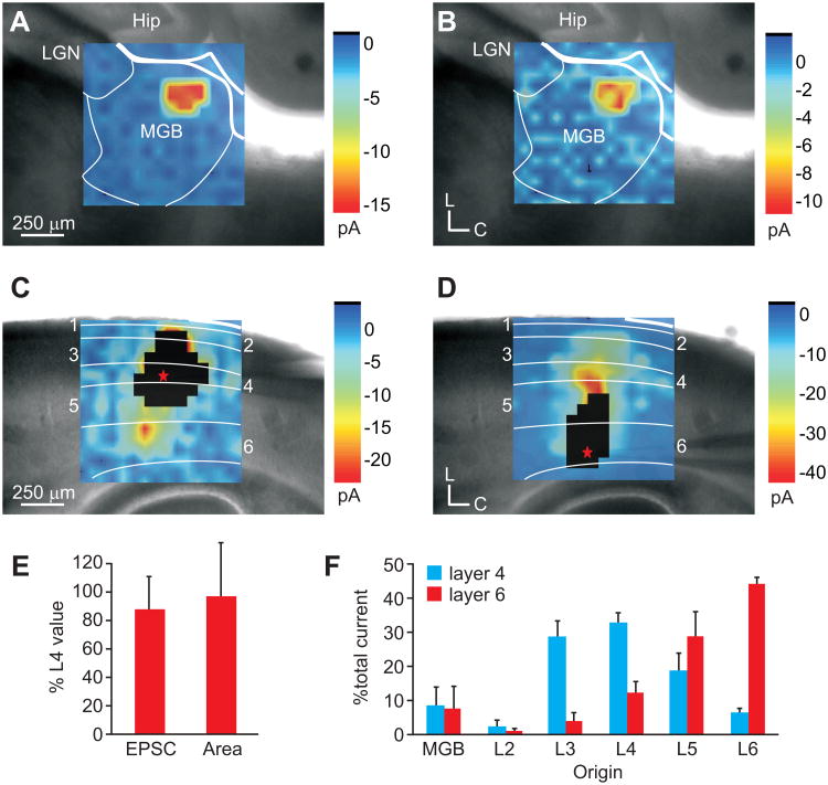
Fig. 5.
Auditory thalamocortical and intracortical inputs to layers 4 and 6 of A1. (A-D) Average LSPS plots of mean EPSCs recorded in a layer 4 neuron (A,C) or a layer 6 neuron (B,D) in response to photostimulation of the medial geniculate body (MGB; A-B) or auditory cortex (A1; C-D). White boxed regions in C and D illustrate direct response areas of recorded neurons. (E) Mean thalamic area and mean total evoked current in layer 6 normalized to that of layer 4 neurons recorded in the same column. (F) Mean percent of total current elicited from the MGB and layers 2-6 in either the layer 4 neuron (black) or layer 6 neuron (grey).

