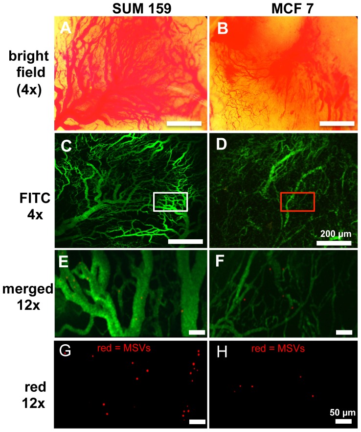
Figure 1. Visualization of tumor particle dynamic flow by intravital microscopy.
A, B) Wide-field images of SUM159 and MCF7 tumors exposed via skin-flap. SUM159 tumors are characterized by highly dense network of dilated vessels whereas MCF7 tumors are mostly comprised of smaller, more widely spaced vessels; C, D) Tumor vasculature was delineated following bolus i.v. injection of 70 kDa FITC-dextran; E-H) At higher magnification, individual rhodamine-labeled MSVs were readily identified. Merged images (E, F) show particle localization relative to the tumor vessels 60 sec after particle injection.

