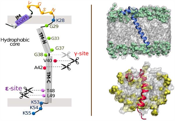
Figure 1.
(Left) Schematic of C99 showing key sequence information, likely secondary structure regions, ∊- and γ- cleavage sites, and approximate insertion within the membrane bilayer. The break in the TM helix at the “GG kink” between G37/G38 is indicated. (Right) Depiction of C9915–55 monomer in a POPC lipid bilayer (above) showing an average tilt angle of 22.5 (deg) with respect to the bilayer normal and (below) C9915–55 in a DPC micelle. The phosphocholine group is shaded green (POPC) or yellow (DPC).

