
Figure 1.
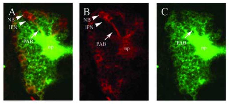
Comparison of labeling with a conventional anti-HRP antibody and with an elav::Gal4 driven UAS::syn-GFP. (A–C): dorsal views of brains of stage 15 embryos carrying the elav::Gal4 driven UAS::syn-GFP inserts, stained with anti-GFP (B; red) and anti-HRP (C; green); (A) is a merge of (B) and (C). Anti-GFP immunostaining (B) shows primary axon bundles (PAB; arrow) and late-born primary neurons (lPN) from which they emanate. Many clusters still include neuroblasts (NB). (C) Anti-HRP antibody immunoreactivity includes the PABs and the embryonic neuropile (np). Since all neuronal cell bodies show strong anti-HRP immunoreactivity, the primary axon bundles and their associated primary cell clusters can not be distinguished very clearly. In contrast, elav::Gal4 driven UAS::Synaptobrevin-GFP shows a marked temporal dynamics in that the more immature neurons of the primary cell clusters, which are located near the cortex surface, express the reporter gene at much higher levels than the more mature neurons and their neurites (Fig. 1B). This makes it possible to anatomically map the primary cell clusters and PABs individually.