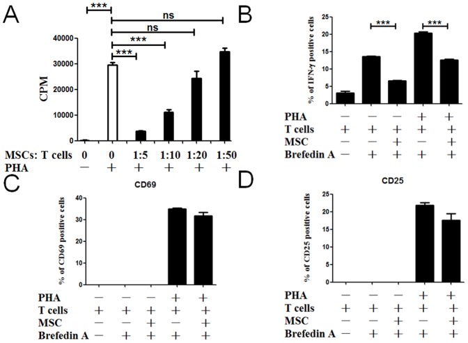
Figure 2. MSCs are capable of suppressing T cells proliferation.
T cells were cocultured with MSCs. All T cells were activated with PHA/IL-2 except control. Different MSCs: T ratios (1∶5, 1∶10, 1∶20 and 1∶50) were used to perform the MLR. T-cell proliferation was assessed by [3H] thymidine incorporation assay after 72 h of culture (A). n = 4. When it says NS, they are being compared to the grey column (MSC 0 PHA +). (B–D): T cells activated with or without PHA/IL-2 were cultured with UCMSCs at MSCs: T ratios (1∶10). After 24 hours, CD8+ cells were analyzed for intracellular IFN-γ and for the expression of activation molecules using anti-CD25 or anti-CD69 monoclonal antibodies. For IFN-γ staining, T cells were treated with Brefeldin A for the last 4 hours of cultures. Cells were permeabilized and the proportion of CD8+/IFN-γ+, CD8+/CD69+ and CD8+/CD25+ T cells was quantified. n = 4. All data are representative of three independent experiments. ***, p<0.001.

