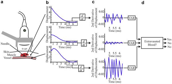
Fig 1.

Methods of blood signal detection in acoustic radiation force impulse (ARFI) ultrasound. (a) The imaging transducer is positioned above punctured muscle and surrounding soft tissue and small (~2 mm) vein. (b) ARFI displacement profiles arising from regions of hemorrhage and tissue (top), muscle and soft tissue alone (middle) and luminal blood (bottom) exhibit late displacement variations (boxed) that are highlighted by (c) the second time derivative. (d) Applying upper and lower thresholds to the variance of the second time derivative of displacement distinguishes hemorrhage from soft tissue and luminal blood.
