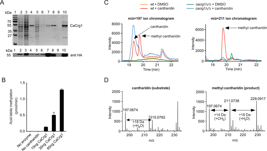
Figure 2. CaCrg1 is a small molecule MTase.
A. Coomassie-stained 12% SDS-PAGE of purified His-tagged CaCrg1. lane 1, molecular weight standards; lane 2, soluble cell extract; lane 3, insoluble fraction; lane 4, Ni2+ Sepharose beads after wash 1; lane 5, unbound to beads cell extract; lane 6, wash 1; lane 7, beads after three washes; lane 8, non-concentrated elute; lane 9, flow-through; lane 10, concentrated and desalted elute. The expression of CaCrg1 was assessed with a mouse monoclonal anti-HA antibody (bottom).
B. CaCrg1 shows robust MTase activity with cantharidin as the substrate in vitro. The reactions containing varying amounts of CaCrg1 enzyme and cantharidin were tested on production acid-labile methylated ester. The error bars represent the standard deviation of two separate experiments each performed in duplicate.
C. CaCrg1 is required for a formation of methyl cantharidin in vivo. Wt and cacrg1Δ/Δ cells were cultured in the presence and absence of cantharidin before extraction of intracellular metabolites and analysis by LC-MS/MS. Single-ion chromatograms of various cellular extracts are shown for the mass ranges corresponding to cantharidin (m/z=197±100 ppm) (left panel) and methyl cantharidin (m/z=211±100 ppm) (right panel). Arrows mark the elution patterns for cantharidin and methyl cantharidin.
D. Averaged spectra of the cantharidin (left panel) and methyl cantharidin (right panel). Chromatographic peaks from the cantharidin-treated wt are shown and ions of interest are indicated.

