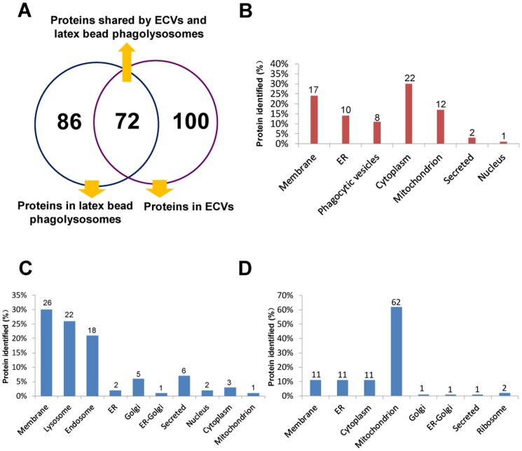
Figure 1. Graphic representation of the proteins identified in ECVs and phagolysosomes containing latex beads.
(A): Schematic representation of proteins present in ECVs and latex bead phagolysosomes; (B): Proteins shared by ECVs and latex bead-containing phagolysosomes were grouped according to their cellular locations; (C): Proteins detected in all latex bead-containing phagolysosomes but not detected in any ECVs were classified based on their cellular locations; (D): Proteins present only in ECVs were grouped according to their cellular locations.

