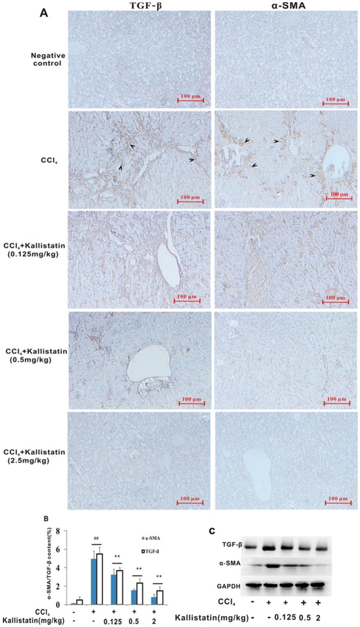
Figure 3. Kallistatin prevents CCl4–induced liver fibrogenesis in rats.
(A) Representative images of immunohistochemical staining for TGF-β1 (brown in color, arrowheaded) and α-SMA (brown in color, arrowheaded) are shown (original magnifications ×10) respectively. Expression of α-SMA around the periportal fibrotic band areas, central vein and fibrous septa were arrowed. Scale bars = 100 µm. (B) Deposition of α-SMA and TGF- β1 was quantitated by image analysis based on the immunohistochemistry results. Data are expressed as mean±SD (n = 8). ## p<0.01 vs. negative control; ** p<0.01 vs. model control group. (C) Immunoblotting analysis of α-SMA and TGF- β1 in livers from CCl4 alone or plus kallistatin treated rats. Data from immunoblotting were confirmed, showing kallistatin-dependent abrogation of α-SMA and TGF-β1 expression. Immunoblotting and immunohistochemistry results were consistent. Housekeeping proteins GAPDH are useful as loading controls for western blot and protein normalization.

