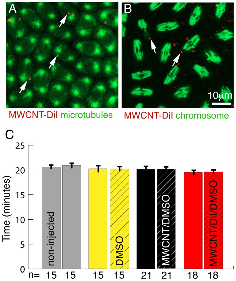
Figure 2. MWCNTs do not interfere with cell division.
Ventral views; Anterior is up. Bar equals 10 µm in A, B. (A) Single live confocal section through the syncytial blastoderm during a division wave. Microtubules are labelled with GFP (green, Jupiter-GFP). All nuclei are in the same stage (metaphase) of the cell cycle even if MWCNTs (red, arrows) are present. (B) Single live confocal section through an embryo with YFP labelled histones marking chromosomes (green). Regardless of the presence or absence of MWCNTs (red, arrows), all nuclei are in the same stage of division (anaphase). (C) The rate of nuclear divisions is not affected by the presence of MWCNTs. Division times for three nuclei per embryo half were recorded and division times between non-injected (non-hatched) and injected (hatched) halves compared. For non-injected embryos (grey) division times of three randomly chosen nuclei for each half were recorded. We detect no differences in the division times between non-injected and injected halves nor between embryos injected with 10% DMSO (yellow, solvent control), 1 mg/ml MWCNT in 10% DMSO/water (black, MWCNT/DMSO) and 1 mg/ml MWCNT in 10% DMSO labelled with DiI (red, MWCNT/DiI/DMSO). Y-axis, division time in minutes; X-axis, Number of nuclei analysed (n).

