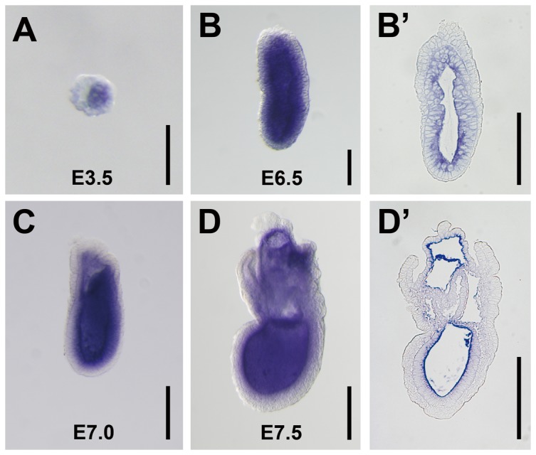
Figure 1. Expression of Meteorin in early embryogenesis.

(A–D) Expression of Meteorin mRNA at different stages of development was analyzed by whole-mount in situ hybridization with embryos from a timed-pregnant mouse. (A) Meteorin was expressed in the inner cell mass at the E3.5 blastocyst stage. (B, B′) At E6.5, the pre-steak stage, Meteorin was expressed in the extra-embryonic ectoderm and epiblast, but not in the visceral endoderm. (C, D, D′) During E7.0–E7.5, the mid- to late-gastrulation stages, Meteorin expression was sustained in extra-embryonic ectoderm and the epiblast, but not in extra-embryonic tissue. (B′ and D′) After in situ hybridization with a probe specific for Meteorin at each developmental stage, embryos were fixed, embedded in paraffin, and then sectioned sagittally. The expression level was relatively high in cells close to the proamniotic cavity. All experiments were conducted with more than 5 embryos at each developmental stage and the representative images were captured. Scale bars in A, B, and B′: 100 µm. Scale bars in C, D, D′: 200 µm.
