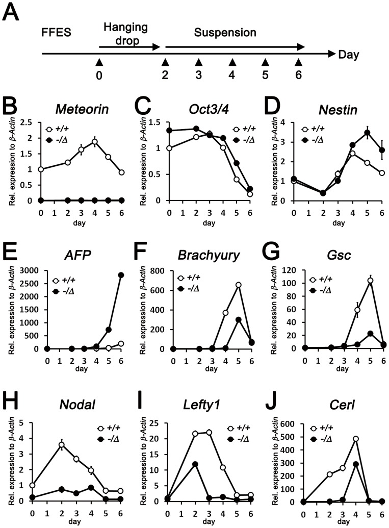
Figure 3. Impaired mesendodermal development in EB culture of Meteorin−/Δ cells.
(A) Schematic view of EB culture. After 2 passages maintained in feeder-free embryonic stem cells, cells were dissociated and cultured using hanging drops for 2 days. Then, they were collected and cultured on bacterial-grade dishes. At each day of culture, indicated as arrowheads in the schematic view, cells were harvested for subsequent qRT-PCR experiments. (B–H) The expression of early developmental lineage markers on each day of culture was analyzed by qRT-PCR. (B) Successful gene deletion in Meteorin−/Δ ES cells was confirmed by observing the transcript level of Meteorin. (C) Expression of Oct3/4, a pluripotency marker, was similarly maintained in Meteorin−/Δ EB culture compared to Meteorin+/+ EB culture. Expression levels of Nestin, an early neuroectoderm marker (D), and AFP, a visceral endoderm marker (E), were higher in Meteorin−/Δ EB culture than in Meteorin+/+ EB culture. The expression levels of Brachyury, an early mesendoderm marker (F), Gsc, an endoderm marker (G), and Nodal, a posterior epiblast marker (H), significantly decreased in Meteorin−/Δ EB culture throughout the culture period. The expression of Lefty1 (I) and Cerl (J), downstream molecules of Nodal signaling, was also lower in Meteorin−/Δ EB culture than in the control. Error bars indicate standard error of the mean (s.e.m.). All experiments were conducted more than 3 times and the representative graphs are shown.

