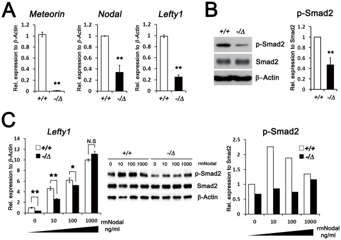
Figure 4. Reduced, but intact, Nodal signal transduction pathway in Meteorin−/Δ ES cells.
(A) Relative levels of Meteorin, Nodal, and Lefty1 in Meteorin+/+ and Meteorin−/Δ ES cells were analyzed by qRT-PCR. In Meteorin−/Δ ES cells, the transcript levels of these genes decreased. Error bars indicate standard deviation (s.d.). **p<0.01. (B) Smad2 and its phosphorylated level (p-Smad2) were investigated by western blotting, and the protein loading was normalized to β-Actin. In Meteorin−/Δ ES cells, the phosphorylated level of Smad2 significantly decreased. Scanned blot images were measured by Scion image. Error bars indicate s.d. **p<0.01. (C) Expression of Lefty1, a downstream molecule of Nodal signaling, was assessed by qRT-PCR at 24 hours after addition of recombinant mouse Nodal (rmNodal) protein to Meteorin+/+ or Meteorin−/Δ ES cells. Levels of Smad2 and p-Smad2 were also measured by western blotting at 2 hours after rmNodal treatment. The reduced levels of Lefty1 transcript and p-Smad2 were rescued by rmNodal treatment in Meteorin−/Δ ES cells. Scanned blot images were measured by Scion image.

