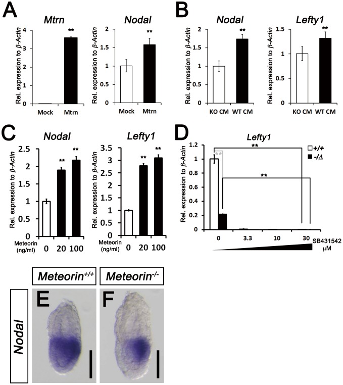
Figure 5. Restoration of Nodal signaling in Meteorin+/Δ ES cells by Meteorin expression and reduced expression of Nodal in Meteorin−/− embryos.
(A) At 48-mMeteorin plasmid into Meteorin−/Δ ES cells, Meteorin and Nodal expression was higher than that of pCAGGS mock-transfected Meteorin−/Δ ES cells. (B) When conditioned medium obtained from Meteorin+/+ ES cells (WT CM) was applied to Meteorin−/Δ ES cells, the expression levels of Nodal and Lefty1 were higher than those seen when conditioned medium from Meteorin−/Δ (KO CM) was applied. (C) Nodal and Lefty1 expressions were analyzed by qRT-PCR 48 hrs after the addition of recombinant mouse Meteorin (rmMeteorin) to Meteorin−/Δ ES cells. Both gene expressions were induced by Meteorin protein addition. Error bars indicate s.e.m. **p<0.01. (D) Expression of Lefty1 transcript was assessed by qRT-PCR after the addition of SB431542, an inhibitor of type I TGF-beta receptors, to Meteorin+/+ or Meteorin−/Δ ES cell lines. The residual activity of TGF-beta/Nodal signaling in Meteorin−/Δ ES cells was further inhibited by SB431542 treatment. Error bars indicate s.e.m. **p<0.01. (E–F) Expression of Nodal transcripts was analyzed by in situ hybridization in Meteorin+/+ and Meteorin−/− embryos at each developmental stage. At E6.5, the expression level of Nodal was significantly lower in Meteorin−/− embryos than in Meteorin+/+ embryos, especially in the proximal epiblast. All scale bars: 100 µm.

