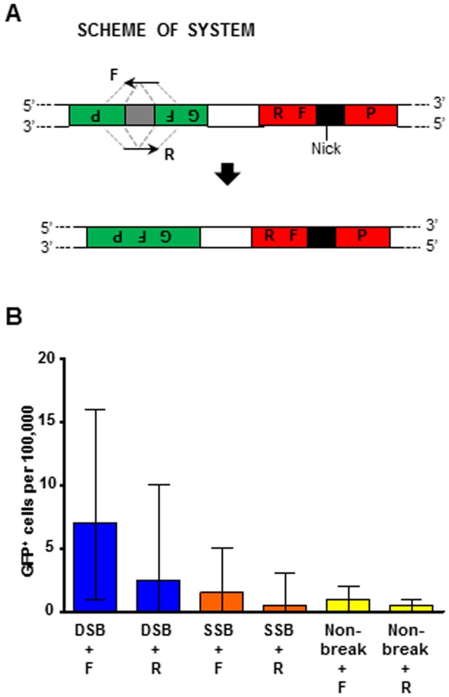
Figure 7. Gene targeting distant from the I-SceI DSB or SSB in human cells.
(A) Scheme showing the disrupted GFP plasmid locus 2.3 kb distant from the I-SceI recognition sequence (black box). The position of the SSB is indicated (“Nick”). Dashed gray lines indicate the complementarity between the F oligonucleotide and the antisense strand of the targeted gene, and between the R oligonucleotide and the sense strand of the targeted gene. (B) Frequencies of GFP+ cells following expression of wild-type I-SceI (dark blue bars labeled “DSB”), K223I I-SceI (orange bars labeled “SSB”), or D145A I-SceI (yellow bars labeled “Non-break”) using either of the single oligonucleotides to correct the GFP gene distant from the I-SceI break. All data are presented as the median with range (n = 6). For the specific numerical values see Table S3K.

